Section III — Transplant Ledger
Transplant Nephrology
Split-view monitoring of immunosuppression and graft health. Glassmorphic containers separate pharmacologic data from graft function metrics for vigilance at every glance.
Immunosuppression
Drug levels & regimens
Tacrolimus (CNI)
Maintenance — Calcineurin inhibitor
Target
Trough 5–12 ng/mL
- Monitor trough levels religiously — narrow therapeutic window
- CYP3A4 interactions: azoles, macrolides ↑ levels; rifampin ↓ levels
- Adverse effects: nephrotoxicity, tremor, hyperglycemia, alopecia
- Do not combine with cyclosporine
Mycophenolate (MMF)
Maintenance — Antimetabolite
Target
500–1000 mg BID
- Inhibits lymphocyte purine synthesis
- Adverse effects: leukopenia, GI upset, teratogenicity
- Monitor CBC monthly — dose reduce if WBC < 3.0
- Avoid in pregnancy — switch to azathioprine
Prednisone
Maintenance — Corticosteroid
Target
Taper to 5 mg daily
- Tapered over months post-transplant
- Long-term: osteoporosis, HTN, hyperglycemia, infection risk
- Some protocols allow steroid withdrawal — higher rejection risk
- Bone density monitoring + calcium/vitamin D supplementation
Induction Therapy
Perioperative — Depletion or blockade
Target
Given at transplant
- Basiliximab: IL-2 receptor blocker — low infection risk
- ATG (rabbit): lymphocyte-depleting — higher-risk recipients
- Used in high immunologic risk or delayed graft function
- Alemtuzumab: profound depletion, single dose
Graft Function
Recipient monitoring metrics
Serum Creatinine
Stable or decreasing
> 25% rise from baseline → suspect rejection
eGFR
≥ 60 mL/min ideal
Sustained decline → evaluate cause
Urine Output
Adequate (> 1 L/day)
Oliguria → assess volume, obstruction, rejection
Proteinuria
UPCR < 0.5 g/g
New proteinuria → consider biopsy
Acute Rejection — Recognition
T-cell Mediated (TCMR)
Days to months
Clinical Signs
Rising SCr, fever, graft tenderness, reduced urine output
Treatment
Pulse steroids (methylpred 500mg IV × 3 days); resistant → ATG
Antibody Mediated (ABMR)
Any time post-transplant
Clinical Signs
Rising SCr + DSA positive; C4d staining on biopsy
Treatment
Plasmapheresis, IVIG, rituximab; bortezomib if severe
Chronic Active ABMR
Months to years
Clinical Signs
Insidious GFR decline, proteinuria, transplant glomerulopathy
Treatment
Optimize immunosuppression; limited treatment options
Allograft Biopsy
Indications, histology & rejection management
When to biopsy
- 1Serum creatinine rise ≥ 25% from baseline (unexplained)
- 2New or worsening proteinuria (UPCR > 0.5 g/g, or new nephrotic-range)
- 3Persistent delayed graft function or oliguria beyond expected recovery
- 4Suspected rejection — fever, graft tenderness, rising SCr — after excluding obstruction & infection
- 5Protocol (surveillance) biopsy at fixed intervals in high-immunologic-risk recipients
- 6Distinguish rejection from CNI nephrotoxicity, recurrent disease, or infection
Histology atlas — normal vs rejection
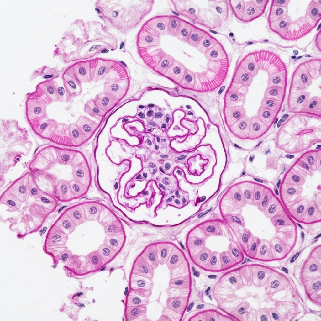
Normal
Normal Allograft
PAS · 400×
- Normal glomerulus — thin capillary loops, open Bowman's space
- Intact tubular epithelium, no tubulitis (t0)
- Minimal interstitial inflammation (i0)
- No C4d staining, no transplant glomerulopathy
No rejection. Continue current immunosuppression; look elsewhere for the cause of dysfunction.
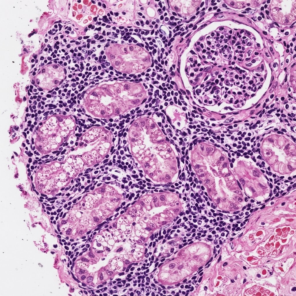
Acute Rejection
Acute T-cell Mediated Rejection
H&E · 400×
- Dense interstitial lymphocytic infiltrate (i2–i3)
- Tubulitis — lymphocytes invade tubular epithelium (t2–t3)
- ± Intimal arteritis (v1–v3) — grades severity
- Glomerulus with mild inflammation
Acute TCMR. Grade by Banff (IA–III) and treat with pulse steroids ± ATG.
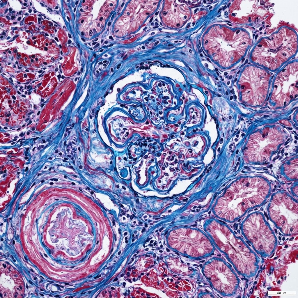
Chronic Rejection
Chronic Active ABMR
Trichrome · 400×
- Transplant glomerulopathy — double-contour (split) GBM (cg)
- Interstitial fibrosis + tubular atrophy (IFTA)
- Peritubular capillary basement membrane multilayering
- ± C4d positive and donor-specific antibody present
Chronic active ABMR. Optimize immunosuppression; limited reversibility; manage comorbidities.
Management by biopsy result
T-cell Mediated Rejection
Steroid-responsive; escalate by Banff grade
Borderline / IA
Methylprednisolone 250–500 mg IV daily × 3 days; optimize tacrolimus trough + MMF; re-biopsy if no response.
IB / IIA
Steroid pulse + consider ATG (1.5 mg/kg/day) if steroid-resistant or severe; increase maintenance immunosuppression.
IIB / III (vascular)
ATG + IV steroids; aggressive inpatient treatment; monitor hemodynamics and graft perfusion.
Antibody-Mediated Rejection
Target the donor-specific antibody
First-line
Plasmapheresis (3–6 sessions) to remove DSA + IVIG (2 g/kg total, divided).
Add-on
Rituximab (anti-CD20) ± bortezomib (proteasome inhibitor) if severe or persistent.
Optimize / refractory
Maximize tacrolimus + MMF; consider eculizumab for complement-mediated refractory disease.
Chronic Active ABMR
Slow the decline; preserve residual function
Immunosuppression
Optimize baseline tacrolimus + MMF; ensure adherence; rituximab + IVIG if DSA-driven active ABMR.
Nephroprotective
Control BP; ACEi/ARB for proteinuria; avoid nephrotoxins; manage cardiovascular risk.
Graft failure
Re-transplant evaluation; dialysis access planning if eGFR declines to ESRD.
Post-Transplant Infection Timeline
1
Month 1
Nosocomial & donor-derived
- Wound infections
- UTI
- C. difficile
- Donor-derived (rare)
2
Months 1–6
Opportunistic infections peak
- CMV (prophylaxis with valganciclovir)
- BK virus nephropathy
- PJP (prophylaxis with TMP-SMX)
- Candida
3
> 6 Months
Community-acquired & chronic
- Community respiratory viruses
- UTI (recurrent)
- Late opportunistic (if prophylaxis stopped)
References & Further Reading
- 01KDIGO Clinical Practice Guideline for the Care of Kidney Transplant Recipients — KDIGO (2009)
- 02Banff Classification of Renal Allograft Pathology — Loupy A et al., Am J Transplant (2023)
- 03AST Infectious Diseases Guidelines for Transplant Recipients — American Society of Transplantation (2019)
- 04KDIGO Consensus Conference on Immunosuppression in Kidney Transplant Recipients — KDIGO (2021)
- 05Management of antibody-mediated rejection in kidney transplant — Eskandary F et al., Transplant Rev (2020)